Treating Equine Lymphangitis
Lymphangitis, a swelling of the lymphatic vessels, generally in the hind limbs, has little scientific research surrounding it and therefore can be difficult to pinpoint and treat. Also, due to the delicate structures of the horse’s distal limbs, a fat leg is a common thing to most equestrians, with there being several different causes; infection, trauma or foot issues being some of the many causes.
Lymphangitis has a tendency, as with most causes of chronic swelling in the distal limb, to reoccur, which makes it a tricky condition to treat.
The lymphatic system is designed to gather liquid from the body through a series of tubes that act like capillaries and return it to the cardiovascular system. Conversely the vessels also act as drains, mopping up water and macromolecules from the blood system. (Sellnow. L, 2000)
When the flow of lymph through the skin is blocked, for whatever reason, the system ‘backs up’ and the interstitial space becomes flooded causing swelling and pain response.
"Lymphangitis is a condition that results in swelling of the leg and is thought to be due to a restriction in lymphatic flow, possibly due to a bacterial infection," Rose and Hodgson write in their Manual of Equine Practice. They go on to say that, "Corynebacterium pseudotuberculosis has been cultured from affected horses, but in many cases bacterial culture will be negative. Lymphangitis sometimes occurs in outbreak form, and we have seen up to 20 horses affected in a mounted police stable. The mode of transmission and the etiologic agent could not be established." (Rose & Hodgson, 1999)
Within the research, it was found near impossible procure adequate samples for bacteriology, making it difficult to find the causative agent.
As with all cases of swelling, cryotherapy and compression help in management of the condition. When the condition progresses to ulcerative lymphangitis, where the lesions happen in the affected area, which often ooze and leak lymph. This is believed to be caused by bacteria entering the skin, so good stable management is very important.
Expediency is also crucial. The vet will usually prescribe anti-inflammatories, steroids and antibiotics. Gentle exercise is also important to increase drainage within the limb. This will often be very painful for the patient, but is necessary.
Laser therapy
Laser or red light therapy has long been used on humans to treat pain and swelling. It is effective to treat equine lymphangitis for many reasons, it can modify the viscosity of the lymph making it easier to ‘flow’, it also reduces the oedema, meaning less pain for the patient and enabling more movement in the effected limb. The blue light therapy is an effect antibacterial modularity, proving to be very useful specifically in the ulcerative lymphangitis, due to the large amounts of open wounds on the effected limb.
Case study 1
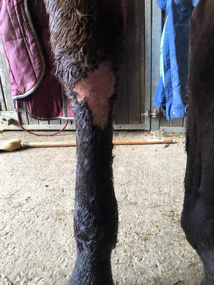
‘Ocean’s Fury’ a 12 year old TB gelding. Has been semi-retired through arthritis of the facet joints of the back. Has history of skin conditions. No lesions found on leg.
Owner found patient with a very filled and swollen leg out of the box in the morning. 5/5 lame on effected limb. Vet attended patient and diagnosed ulcerated lymphangitis. Antibiotics and pain relief were prescribed. Owner advised to walk patient for 5 minutes twice a day.
I attended patient the day after the veterinary diagnosis. Owner had cleaned and massaged limb, removing any dead skin and excess hair. Red light therapy was applied over the entire limb, then specific attention given around the periphery of the ulcerated wounds. Treatment time was 15 minutes in total.
Blue light therapy was also applied over the ulcers to inhibit bacterial proliferation.
The patient then received 15 minutes of red light therapy and 5 minutes of blue light every day for 5 days, alongside his veterinary treatment.
Treatment outcome
After 5 sessions of red light therapy, the hair and infected skin started to slough away, revealing healthy pink skin underneath. After veterinary inspection, it was declared the horse was healing exceptionally well. The lymph was draining through the skin and the oedema had lessened. The horse was visibly less lame.
10 days post diagnosis, horse is now being turned out on hard standing daily and is 2/5 lame, with minimal swelling in the affected limb. The skin has healed beautifully and the vet and owner are delighted with the outcome.
Addendum
The limb, although ‘clean’ with good hair regrowth, suffered a set-back with the oedema returning. I revisited and increased the length of treatment. Patient is now being ridden to assist in in drainage of the limb, which works well. I have also suggested compression bandages on the effected limb at night.
Conclusion
Equine lymphangitis is a pervasive and reoccurring condition, for reasons that are still to be fully researched. However, this small study, and other peer-reviewed papers confirm the use of red light therapy to assist in the healing of all skin conditions.
I have experienced excellent result using this modality for this condition. The treated skin regenerated far faster than untreated skin I’ve witnessed previously and the vet was impressed with how quickly the infected skin sloughed away and healthy skin showed underneath. Upon treatment conclusion, there was new hair growth coming through the worst affected areas.
